Scientific Breakthrough in Medical AI: AIUB-Led Team’s Interpretable Deep Learning Framework for Breast Cancer Diagnosis Published in Q1 Elsevier Journal
Breakthrough Deep Learning System Combines High Accuracy with Transparency, Marking Significant Step Toward Trustworthy AI in Medical Imaging
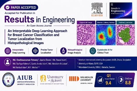
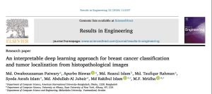
Dhaka, Bangladesh: In a landmark achievement for Bangladeshi artificial intelligence research, a multidisciplinary team led by American International University Bangladesh (AIUB) has developed an innovative deep learning framework that not only detects breast cancer from tissue samples with remarkable accuracy but also explains its diagnostic reasoning, addressing one of the most critical barriers to clinical adoption of AI in healthcare. The study, titled “An Interpretable Deep Learning Approach for Breast Cancer Classification and Tumor Localization from Histopathological Images,” was published online on July 21, 2026, in Results in Engineering (Volume 32, Article 112107), a prestigious journal under the Elsevier publishing umbrella. The journal holds a 2026 Impact Factor of 9.4 , a CiteScore of 8.8, an SJR of 1.240, and is ranked in the Q1 category for General Engineering . The publication represents a significant milestone for AIUB’s growing reputation in computational research and underscores Bangladesh’s emerging role in global medical AI innovation. The paper is available online at DOI: https://doi.org/10.1016/j.rineng.2026.112107.
Md. Owafeeuzzaman Patwary
The research was led by Md. Owafeeuzzaman Patwary, a computer science researcher at AIUB whose work focuses on AI applications in medical imaging and healthcare diagnostics. Owafee, as he is known among his peers, conceptualized the study from its very inception, designed the comprehensive methodology, conducted the formal analysis and investigation, and wrote the original manuscript. His passion for making AI more transparent and trustworthy in medical contexts drove the team’s emphasis on explainability, ensuring that the framework not only delivers accurate classifications but also provides visual insights into how those decisions are made. Owafee’s vision extends beyond this single publication, as he is deeply committed to advancing interpretable AI systems for healthcare, particularly in resource constrained settings where access to expert pathologists may be limited. This research represents his own original contribution to the field, reflecting his unique approach to blending cutting edge deep learning architectures with practical clinical needs. His work demonstrates a rare combination of technical expertise in state of the art neural networks and a deep understanding of the real world constraints facing pathologists, making the framework both innovative and grounded in clinical reality.
He was joined by fellow AIUB researchers Apurbo Biswas, Md. Taufiqur Rahman, and Syeda Asrafa Islam, who contributed across data curation, formal analysis, visualization, validation, and manuscript preparation. Among them, Apurbo Biswas put in tremendous hard work throughout the project, playing a pivotal role in implementing the experimental framework, running extensive simulations, and ensuring the reproducibility of the results. His dedication and meticulous attention to detail were instrumental in validating the model’s performance across multiple evaluation metrics.

Md. Reazul Islam
The collaboration also included Md. Reazul Islam from the University at Albany, State University of New York, where he is currently pursuing his PhD and serving as a Graduate Teaching Assistant, while also holding a position as Assistant Professor at AIUB. He served as project administrator, providing strategic guidance and oversight throughout the research process. His experience and insights helped shape the direction of the study and ensured that the work adhered to rigorous academic standards.

Prof. Dr. Mohammad Firoz Mridha
The project was supervised by Prof. Dr. Mohammad Firoz Mridha, Professor and Head of the Department of Computer Science at AIUB, who served as the corresponding author alongside Md. Rakibul Islam. Dr. Mridha guided the research from its inception, shaping the methodology, providing strategic oversight, and overseeing the manuscript review and editing process. His extensive expertise in artificial intelligence, machine learning, and deep learning applications has been instrumental in establishing AIUB as a hub for cutting-edge research in Bangladesh. Dr. Mridha has been recognized in the prestigious Stanford University and Elsevier’s Top 2% Scientists Worldwide list, an independent global ranking based on citation impact that identifies the most influential researchers across all scientific disciplines. His leadership and mentorship have created an environment where early-career researchers like Owafee can pursue ambitious projects with international impact.
Dr. Md. Abdullah Al Jubair
Dr. Md. Abdullah Al Jubair, Associate Professor at AIUB, provided guidance throughout the research, contributing his expertise in machine learning, computer vision, and biomedical applications. His extensive experience in deep learning architectures and medical image analysis proved invaluable in refining the ensemble framework and ensuring its clinical relevance. As a senior faculty member, Dr. Al Jubair has been instrumental in mentoring early-career researchers and fostering a culture of innovation within AIUB’s Department of Computer Science.
Md. Rakibul Islam from Mälardalen University in Sweden served as corresponding author, contributing to manuscript review, validation, and funding acquisition. His involvement ensured that the research met rigorous international academic standards and facilitated the global reach of this collaborative work. His contributions also helped secure the necessary resources and institutional support that made this multi-national research endeavor possible.
Breast cancer diagnosis today continues to rely heavily on pathologists examining hematoxylin and eosin stained tissue slides under a microscope, a process that, while accurate in expert hands, remains labor intensive, time consuming, and subject to variability between different reviewers. With global breast cancer caseloads continuing to rise, researchers have increasingly turned to deep learning to help automate and enhance parts of this analysis. However, prior AI systems in this space have often struggled with three critical issues simultaneously: data leakage risks where images from the same patient appear in both training and test sets, the black box problem where neural networks cannot explain why they reached a particular conclusion, and weak localization that typically requires pathologists to manually outline tumor regions in advance. The AIUB led team addressed these challenges head on, developing a system that delivers strong classification performance while providing visual explanations and approximate tumor localization, all without requiring expert annotations.
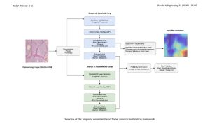
To build their framework, the team created an ensemble system that runs two complementary convolutional neural networks side by side on each image: ConvNeXt Tiny, a modern architecture known for capturing fine grained morphological detail in tissue samples, and MobileNetV3 Large, a lightweight efficient network tuned for high sensitivity. Each network independently estimates the probability that a tissue sample is benign or malignant, and the two probability scores are then averaged together, a probability level fusion approach that improves reliability without adding extra trainable parameters or design complexity. The ensemble strategy was specifically designed to reduce false negatives, cases where a malignant sample is incorrectly labeled benign, because in cancer screening a missed malignant case can delay diagnosis and treatment. The framework was trained and tested on the publicly available BreaKHis dataset, containing 7,909 histopathological images collected from 82 patients at four microscope magnification levels. Critically, the team enforced a strict patient level split between training and testing data, meaning no images from the same patient appeared in both sets, representing a gold standard safeguard against the kind of data leakage that has inflated performance claims in some earlier studies.
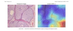
Under this rigorous patient level evaluation protocol, the ensemble model achieved outstanding results with 98.15% accuracy, 98.19% precision, 99.66% recall, 98.66% F1 score, and 99.67% ROC AUC. The exceptionally high recall rate of 99.66% was a particular point of emphasis, as the authors deliberately designed their ensemble strategy to minimize false negatives while maintaining balanced performance across all major metrics. A central feature of the framework is its use of Grad CAM++, an advanced explainable AI technique that generates visual heatmaps showing which regions of a tissue image most influenced the model’s prediction. Rather than treating the neural network as an opaque black box, Grad CAM++ overlays a color coded map on each image, highlighting the areas the model effectively looked at when making a diagnosis. According to the paper, in malignant samples the highlighted regions were consistently associated with dense cellular structures, irregular nuclear shapes, and disrupted tissue organization, features that align with what pathologists already look for, while in benign samples the model’s attention was more diffuse and spread across relatively uniform tissue.
Beyond classification, the team explored whether the same Grad CAM++ heatmaps could be repurposed to approximate where a tumor is located without requiring experts to manually trace tumor boundaries on thousands of images. Using the heatmaps as pseudo labels, they trained a U Net segmentation model under what is known as weak supervision. This localization component achieved a Dice score of 0.5169 and an Intersection over Union score of 0.3519 on the test set. The authors describe this as moderate, rather than clinical grade, segmentation performance, an expected outcome given that the model was trained on approximate, AI generated location cues rather than precise expert annotations. They note explicitly that this part of the framework requires further refinement and is not intended to represent finished, deployment ready tumor mapping. The Grad CAM++ visualizations are framed as an interpretability aid rather than a diagnostic tool in its own right, offering a way to audit and build trust in what the underlying classifier is doing without replacing expert pathological review.
The study drew contributions from researchers across several institutions, with the work carried out within AIUB’s Department of Computer Science and reflecting a multi institutional collaboration spanning Bangladesh, the United States, and Sweden. The authors are direct about the boundaries of their findings, emphasizing that the evaluation relied on a single public dataset and did not include external validation on independent datasets such as BACH or Camelyon16. The study also did not perform statistical hypothesis testing or confidence interval estimation, following an evaluation protocol common in prior BreaKHis based research, meaning the reported metrics should be read as descriptive of this specific test set rather than statistically validated in the broader sense. The paper explicitly states that its results should be interpreted within the scope of controlled experimental conditions, not as evidence of readiness for clinical deployment. The authors call for future work involving multi institutional and cross dataset validation, along with more robust weakly supervised localization strategies, before the approach could be considered for real world diagnostic use.
The framework developed by Owafee and his colleagues brings together three elements, ensemble classification, visual explainability, and approximate tumor localization, that are more commonly studied in isolation. The result, evaluated under a leakage resistant patient level protocol, is a system that performs strongly on the BreaKHis benchmark while offering a degree of transparency into its own decision making. For Bangladesh, this research represents an important step in establishing the country as a contributor to global medical AI innovation, with a growing tech sector and expanding university research capacity positioning the nation to play a meaningful role in developing AI solutions for pressing healthcare challenges. The team’s emphasis on interpretability speaks to a broader trend in AI research: the recognition that high accuracy alone is insufficient for real world medical applications, as clinicians, patients, and regulators increasingly demand that AI systems demonstrate transparency, accountability, and reliability. The authors position their work as a step toward more interpretable and reproducible AI tools for digital pathology research, while emphasizing that substantial further validation stands between this research prototype and any future clinical application.
The authors gratefully acknowledge American International University Bangladesh (AIUB) for providing institutional support and a conducive research environment. The research also received computational resources and research infrastructure support from AMIR Lab (Advanced Machine Intelligence Research Lab). The collaborative contributions of AIUB in Bangladesh, the University at Albany in the United States, and Mälardalen University in Sweden are deeply appreciated. The authors also thank the broader research community for making publicly available datasets such as BreaKHis, which served as the foundation for this study, and acknowledge all patients who contributed to the original data collection.
Reference: Patwary, M.O., Biswas, A., Islam, M.R., Rahman, M.T., Islam, S.A., Jubair, M.A.A., Islam, M.R., & Mridha, M.F. (2026). An interpretable deep learning approach for breast cancer classification and tumor localization from histopathological images. Results in Engineering, 32, 112107. DOI: https://doi.org/10.1016/j.rineng.2026.112107. Published under a CC BY 4.0 license.